Research work on Medical Image Analysis
Medical Image Segmentation Tool (MIST)
Abstract
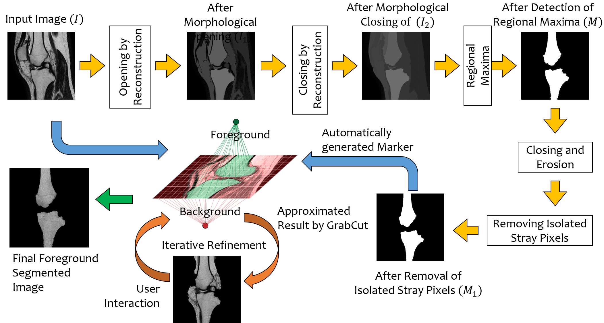
In order to obtain the most suitable method for medical image segmentation, we propose MIST (Medical Image Segmentation Tool), a two stage algorithm. The first stage automatically generates a binary marker image of the region of interest using mathematical morphology. This marker serves as the mask image for the second stage which uses GrabCut to yield an efficient segmented result. The obtained result can be further refined by user interaction.
Published in Computers in Biology and Medicine, Volume 83, pp 22-33, 2017. Available online
Framework
Segmentation is often performed on medical images for identifying diseases in clinical evaluation. Hence it has become one of the major research areas. Conventional image segmentation techniques are unable to provide satisfactory segmentation results for medical images as they contain irregularities. They need to be pre-processed before segmentation. In order to obtain the most suitable method for medical image segmentation, we propose MIST (Medical Image Segmentation Tool), a two stage algorithm. The first stage automatically generates a binary marker image of the region of interest using mathematical morphology. This marker serves as the mask image for the second stage which uses GrabCut to yield an efficient segmented result. The obtained result can be further refined by user interaction, which can be done using the proposed Graphical User Interface (GUI). Experimental results show that the proposed method is accurate and provides satisfactory segmentation results with minimum user interaction on medical as well as natural images.
Screencast of the MIST GUI
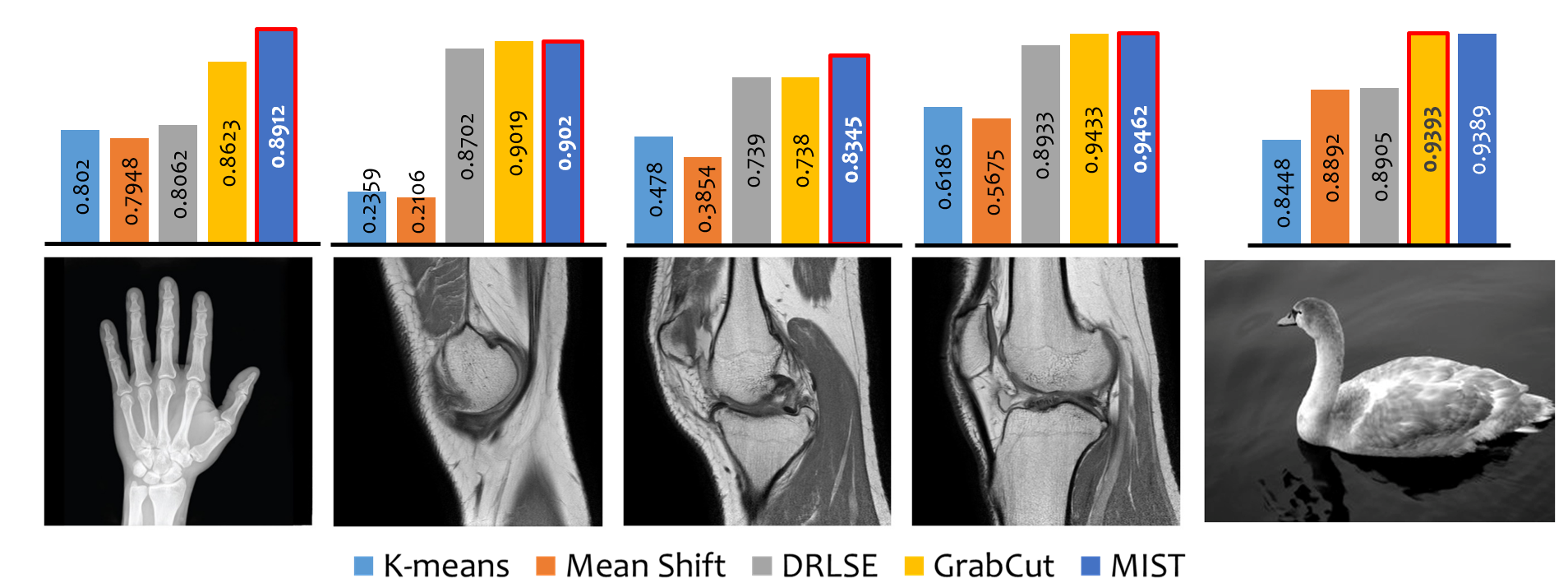
Results
The medical images used in the dataset consist of images in Digital Imaging and Communication in Medicine (DICOM) format. In the dataset used for experiments, there are mainly MRI and X-Ray images of different anatomical regions. Also, natural images have been included in the dataset to check the efficiency of MIST in other domains. The MR images in the data set consist of knee images and are provided by All India Institute of Medical Sciences, Jodhpur, India. The MR images are of size 512 X 512 pixels and their intensity is measured in 16 bits. The X-Ray image dataset is a set of images of different anatomical regions and is a collection of images from the internet. The code is written in MATLAB and C++ using MEX compiler.